


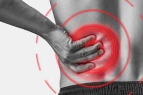
The lumbar region osteochondrosis of the lumbar spine is a chronic degenerative-dynist disease that affects the structure of the intervertebral discs and many lumbar vertebrae.This affects the predominantly working people.It manifests itself in various symptoms with the main back and leg pain, limiting the lower part movement.Diagnostics are used research methods such as radiography, computer tomography, or magnetic resonance of the lumbar spine.In this article you can detail in more detail with the causes, symptoms and methods of osteochondrosis of the lumbar spine.
Osteochondrosis is the result of body aging.These or other signs of the disease are found in almost every person (!) From the age of 25.But here is the severity of these changes, the pace of their progression, and the degree of clinical manifestations depends on many reasons, primarily on how a healthy lifestyle leads to a particular person.Moderate physical activity, mandatory morning gymnastics, body better pose when performing many work (garden, construction, banal cleaning of the house and so on), the orthopedic mattress is the moments that prevent lumbar spine osteochondrosis.
According to statistics, spine osteochondrosis is caused by back pain in 80% of cases.
How does osteochondrosis develop?
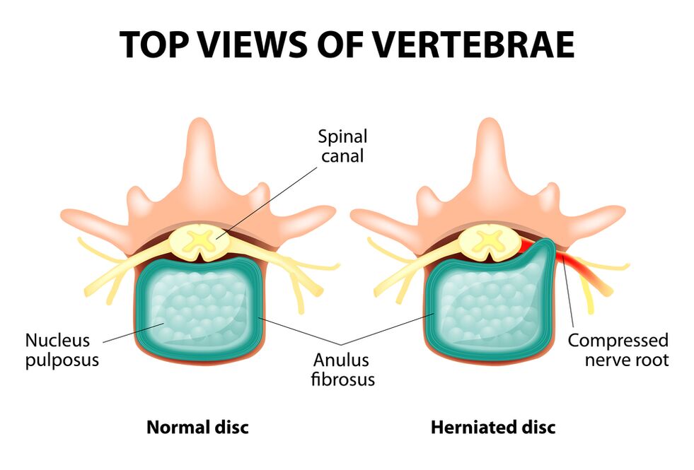
The entire spine consists of separate vertebrae with vertebrae between its bodies.That is, there is a plate between the two vertebrae.The plate consists of a gelatin (pulpic) seed and a fibrous ring.The seed contains a lot of water and provides the spine depreciation and elasticity.The fibrous ring is located along the periphery of the jacket, as if it were holding on its own.
With an increased load of the pier, it changes its physiological properties, loses water and dries, and finally sequences: the plate is flattened and the spinal bodies are close to each other.In addition to such processes, the fibrous ring in the core coat loses its elasticity and extends under the influence of mechanical loads.This is called protrusion.Then the fibrous ring cracks and a gelatin core fall through the gaps obtained: the plate of the plate occurs.Two adjacent vertebrae and the disc between them, the diagram of the spine segment, obtain excessive mobility, thereby increasing the load on nearby segments.The overload of adjacent segments provokes a similar pathological process.These changes are called osteochondrosis.
In order to provide some way, the bone growth is formed along the edge of the spinal bodies, increasing the area of the holder.This phenomenon is called spondylosis.Changes in the joints between the vertebrae are called spondylo arthrosis.Usually, all three abnormal - osteochondrosis, spondylosis, spondyl arthrosis - walk around.
Reasons
Why does osteochondrosis occur?To date, there are many theories:
- Mechanical theory: Perhaps the main cause of the spine should be considered as a regular increase in the spine.This is the reason why osteochondrosis is an almost mandatory fate of movers, miners, builders and people of such professions.The incidence of osteocondrosis of the lumbar region is primarily related to slopes and severity, forced to work uncomfortable;
- Another factor of development is incorrect posture, sitting in a bad pose, which is particularly relevant to mental workers;
- Occasionally, the role plays the role of the hereditary properties of the spine and the nutrition of each structure;
- Traumatic theory: any trauma of the spine (even the most significant) can initiate a degenerative process;
- Hormonal metabolic disorders and endocrine diseases can adversely affect the metabolism of the spinal column tissues and contribute to the development of osteochondrosis;
- Age theory means the natural wear of discs in the process of life.
Rarely, only one of these theories explains the occurrence of osteochondrosis in each case.At the same time, many factors are more often "blamed".
When the lumbar spine occurs in osteocondrosis, the overweight plays an important role as it is overloading the spinal column itself.The higher the body weight index (the degree of obesity), the more the changes in the spine are.Other reasons for provoking the appearance of osteochondrosis can be noted:
- sedentary lifestyle;
- I am, amended, nutrition (fast food, excess sweet, semi -developed products: all this leads to imbalance of trace elements) and lack of fluid;
- Disorders of the spine structure (for example, the presence of an additional lumbar vertebrae);
- Constant wearing high rodent shoes;
- pregnancy (due to excessive load on the lumbar spine);
- Sudden termination of training in sports from a professional point of view;
- Smoking and abuse of alcohol: Like factors that accelerate the aging process in the body.
Symptoms
The main manifestation of osteochondrosis of the lumbar spine is pain.The nature of the pain, the place of occurrence and the direction of distribution depends on which receptors are irritated, that is, the rough changes in the plate and in the surrounding tissues, protrusion or hernia in which the protruding and so on.
Reflex and compression syndromes are distinguished from osteochondrosis of the lumbar spine.
Reflex syndromes develop when the receptors of the affected disk, the ligaments and the joint capsules nearby are irritated.They are reflexive because, in addition to the pain, it is accompanied by muscle-tonic, vegetative-vascular or neurodistrophic reflex changes, ie irritation with reflexes is in other structures and the symptoms are mainly caused by soft tissue side.
Compression syndromes occur as a result of compression (compression) of nerve roots, blood vessels or spinal cord, which is created during osteochondrosis.

Reflex syndrome of lumbar spine
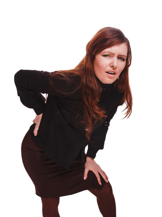
Lumbago(Feeling): acute sudden pain in the lower part, which is unpleasant or physical tension (much less often - for no obvious reason).It is believed that the occurrence of lumbago is related to the movement of the coat seed in the fibrous ring, ie it develops in the initial stages of osteochondrosis.Pain is often called "feeling", "stakes stuck in the lower back."Patients freeze in the pose in which the pain caught them.The smallest step causes an increase in pain (sneezing, coughing, turning to bed, moving the leg).If a person was prone to the development of Lumbago (which is most commonly done), he or she will not be able to compensate.The muscle tension in the lumbar spine occurs reflectively.Along the vertebrae, there is a muscle cylinder in this area that occasionally visible to the naked eye and the muscle tension can be pronounced so much.He feels painful for the patient.Such an increased muscle tone plays an immobilizing role, protecting the affected lumbar segment from pathological mobility, which can trigger the state's deterioration.Natural bends of the spinal column are flat (lordosis) flat (lordosis), possibly curvature (skoliosis) due to muscle tension.
Lumbalgia- The lumbar level is another reflex syndrome.This term also means that pain is present in the lumbar region.Unlike Lumbago, the pain is not acute, but gradually within a few hours or even days.The pain increases stupid, moderate intensity, in motion, in a seat or standing position as it moves from one position to another.A little relief brings to bed or back under the lower part, but passive rise in the straightened leg causes increased pain in the lower back (slow symptom).The palpation of the lumbar spine is painful, but the reflex tension of the muscles is less prominent than with Lumbago and sometimes missing at all.The movement of the lumbar spine is limited but possible.This means that the patient can bend to a certain level and can sideways (then the pain increases).
Sciatica- Another type of lumbar level is reflex syndrome.With this term, they are the pain on the lower back, which gives the buttocks and legs (on the back surface).The pain is different, mostly painful, but can be regularly enhanced with the type of "fireplace" on the foot.As with lumbalgia, it decreases with all movement, walking, tension, lying on the back.The symptom of slow is generally positive.The palpation of the lumbar spine is painful and pushes some points (for example, in the middle of the line that separates the weapon from the thigh, in the middle of the back of the thigh, in the middle of the popliteal fossa).The lower back of the muscles has tension.The predisposition to the pages is limited.
The compression syndromes of the lumbar spine
The clinical property depends on which structure should be compression.
Between the vertebrae in the holes of each vertebrae, nerve roots (spine nerves): left and right.If the pathological formations of osteochondrosis of the lumbar spine (primarily discs of the discs) displace the roots, then the radiculopathy develops, the symptoms of which differ in each root.In all radiculopathics of the lumbar region, the increase in pain is sneezing, coughing, lower back (especially with forward decision), presence of muscle tension in the lower part, restricting movement in the lumbar spine.The following types of lumbar spine are most common:
- Radiculopathy L1, L2, L3: The pain occurs at the lower back, specify the expected thigh.In the same area, the occurrence of paresthesia (feeling of climbing goose bumps, numbness) is possible, the superficial sensitivity disorder (the usual acute touch is not different, cold and hot) are lost.The knee reflex is reduced, the square is explored;
- Radiculopathy L4: In the lower back, the pain in the front of the thigh, slightly lower to the inner surface of the knee joint and the inner surface of the lower leg.In the same areas, paresthesia is felt and the sensitivity of the surface is lost (decreased).The weakness of the thigh quadruple muscles also develops and the knee reflex decreases;
- Radiculopathy L5: One of the common localizations.The pain along the outer edge of the buttocks, along the front of the lower leg to the inner edge of the leg and thumb.Paresthesia can be felt here, superficial sensitivity is disturbed, and causes pain in pain and cough.In addition, it is difficult to extend the thumb of the leg, as the muscle performing this operation is innervated by the Kine L5.Occasionally it is difficult to stand on a corner with one exposed legs;
- S1 radiculopathy is often found with osteochondrosis of the lumbar spine.The pain is the bottom, along the outer edge of the thigh, along the outer edge of the lower leg, to the outer edge of the foot and towards the heel 5.These zones are characterized by a feeling of paraesthesia and a reduction in the sensitivity of the surface.Achilles reflex decreases.Damage to the spine develops the weakness of the lower leg and foot flexors muscles, making it difficult to stand and walk in the socks.
It is possible to develop the radiculopathy of several roots, which is especially characteristic of L5, S1.Sometimes one of the hernia squeezes more roots.
If the disc retires, it can press the spinal cord.This is only possible if the hernia is localized in the upper reference point, since II.There is no spinal cord under the lumbar vertebra (the spinal roots are compressed and the horse tail syndrome is formed).
If the lumbar region's vessels are squeezed into the spinal cord, the spinal cord develops in acute circulatory disorder and longer compression - myelopathy.Myelopathy manifests itself with the bilateral weakness of the leg muscles, starting from the leg and gradually progressing.The sensitivity of the legs is confused, the Achilles reflex is lost and later knees.It is possible to develop urinary disorders (frequent, "imperative" urgency, immediate satisfaction, urinary tract incontinence).
Diagnostic methods
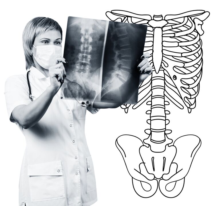
Diagnosis of osteochondrosis of the lumbar spine is based on clinical data and data from further research methods.The key role belongs to methods such as:
- Radiography of the lumbar spine;
- Computer tomography of the lumbar spine;
- Magnetic resonance tomography of the lumbar spine.
The radiography of the lumbar spine is necessarily performed in 2 mutually perpendicular projections-the straight back and the side.Such images allow the shape, contours and structure of the spinal bodies, the height and shape of the intervertebral discs, the spinal disorders and the natural bends.For the display of vertebrae and intervertebral holes, the radiograms are made in oblique projections.In order to identify the pathological mobility of each lumbar segments (a sign of osteochondrosis), radiography is performed in the functional examination, ie in the flexion and extension of the spine.Generally, you can clearly see the height of the vertebral discs in the anterior or back section according to the direction of body bending, due to the functional block of one segment, with osteochondrosis, the height of the disk does not change either during bending or overtime.Pathological mobility determines the displacement of the vertebrae forward or backward.The main X -radiation signs of osteochondrosis include narrowing of intervertebral gap, abnormal mobility and displacement of spinal bodies, salts in the disc (calcification), the development of regional growth of spinal bodies, and the submission of vertebrates from the affected plate (alchondral sclerosis).The radiography of the lumbar spine is the routine of research, which gradually loses its importance to the background of the active implementation of new and informative research methods (CT and MRI).The radiography of the lumbar class is today used as a filtering method for filtering.
The lumbar spine CT is also done using X -Gay radiation, but the radial load of the body is much less than X -Gay.The test is done on a special device desk - a computer tomograph, which is absolutely painless.The resulting images are processed with a computer and allow you to see significantly more structures than the spinal radiography.
MRI is a method in which electromagnetic radiation is used to create images.The examination is also performed in a position on the table, which calls for the tomograph chamber.MRI is harmless and painless.
The lumbar spine CT or MRI allows all the structure of the spine, can carefully examine the intervertebral discs (and the jacket and the fibrous ring), as well as the intervertebral holes, the contents of the spinal canal.Even the mild protrusion of the intervertebral disk is not unnoticed.These methods (especially the MRI) allow them to determine the direction of the disk's hernia, if any, the compression rate of the nerve roots, the spinal cord.Thus, these research methods are much more informative in diagnosis of lumbar spine osteochondrosis than radiography.In addition, they allow not only osteochondrosis but also other diseases (tumors, circulatory disorders in the spinal cord, congenital defects in the spinal structure and spinal cord structure), which is important in differential diagnosis of the back pain.
Lumbar spine osteochondrosis is a disease that most often causes back pain.In fact, this is the destruction of intervertebral discs.Due to osteochondrosis of the lumbar spine, a person often loses working capacity because of the pain in the disease can lead to violation of the mobility of the spine, the inability of seat, position and walking.The symptoms of this disease are not specific and require additional research methods to accurately confirm the diagnosis.The MRI of the spine is the most informative and safe to diagnose osteochondrosis.